Brown Engineering

  • Subscribe to our RSS feed.
  • Twitter
  • StumbleUpon
  • Reddit
  • Facebook
  • Digg
Showing posts with label nano. Show all posts

Thursday, 21 June 2012

Selenium controls staph on implant material

Posted on 08:43 by Unknown
A coating of selenium nanoparticles significantly reduces the growth of Staphylococcus aureus on polycarbonate, a material common in implanted devices such as catheters and endotracheal tubes, engineers at Brown University report in a new study.

PROVIDENCE, R.I. [Brown University] — Selenium is an inexpensive element that naturally belongs in the body. It is also known to combat bacteria. Still, it had not been tried as an antibiotic coating on a medical device material. In a new study, Brown University engineers report that when they used selenium nanoparticles to coat polycarbonate, the material of catheters and endotracheal tubes, the results were significant reductions in cultured populations of Staphylococcus aureus bacteria, sometimes by as much as 90 percent.

Selenium solutionQi Wang swirls a solution of selenium nanoparticles in the lab.
Coatings of the nanoparticles appear effective in fighting staph
bacteria in medical device materials, according to a new study.

Credit: Webster Lab/Brown University
“We want to keep the bacteria from generating a biofilm,” said Thomas Webster, professor of engineering and orthopaedics, who studies how nanotechnology can improve medical implants. He is the senior author of the paper, published online this week in the Journal of Biomedical Materials Research A.

Biofilms are notoriously tough colonies of bacteria to treat because they are often able to resist antibiotic drugs.

“The longer we can delay or inhibit completely the formation of these colonies, the more likely your immune system will clear them,” Webster said. “Putting selenium on there could buy more time to keep an endotracheal tube in a patient.”

Meanwhile, Webster said, because selenium is actually a recommended nutrient, it should be harmless in the body at the concentrations found in the coatings. Also, it is much less expensive than silver, a less biocompatible material that is the current state of the art for antibacterial medical device coatings.

Webster has been investigating selenium nanoparticles for years, mostly for their possible anticancer effects. As he began to look at their antibiotic properties, he consulted with Hasbro Children’s Hospital pediatrician Keiko Tarquinio, assistant professor of pediatrics, who has been eager to find ways to reduce biofilms on implants.

Studying selenium

For this study, Webster and first author Qi Wang grew selenium nanoparticles of two different size ranges and then used solutions of them to coat pieces of polycarbonate using a quick, simple process. On some of the polycarbonate, they then applied and ripped off tape not only to test the durability of the coatings but also to see how a degraded concentration of selenium would perform against bacteria.

On coated polycarbonate — both the originally coated and the tape-tested pieces — Wang and Webster used electron and atomic force microscopes to measure the concentration of nanoparticles and how much surface area of selenium was exposed to interact with bacteria.

One of their findings was that after the tape test, smaller nanoparticles adhered better to the polycarbonate than larger ones.

Then they were ready for the key step: experiments that exposed cultured staph bacteria to polycarbonate pieces, some of which were left uncoated as controls. Among the coated pieces, some had the larger nanoparticles and some had the smaller ones. Some from each of those groups had been degraded by the tape, and others had not.

All four types of selenium coatings proved effective in reducing staph populations after 24, 48, and 72 hours compared to the uncoated controls. The most potent effects — reductions larger than 90 percent after 24 hours and as much as 85 percent after 72 hours — came from coatings of either particle size range that had not been degraded by the tape. Among those coatings that had been subjected to the tape test, the smaller nanoparticle coatings proved more effective.

Staph populations exposed to any of the coated polycarbonate pieces peaked at the 48-hour timeframe, perhaps because that is when the bacteria could take fullest advantage of the in vitro culture medium. But levels always fell back dramatically by 72 hours.

The next step, Webster said, is to begin testing in animals. Such in vivo experiments, he said, will test the selenium coatings in a context where the bacteria have more available food but will also face an immune system response.

The results may ultimately have commercial relevance. Former graduate students developed a business plan for the selenium nanoparticle coatings while in school and have since licensed the technology from Brown for their company, Axena Technologies.
Read More
Posted in axena, biofilm, nano, nanoparticles, selenium, wang, webster | No comments

Monday, 11 June 2012

Small Wonder

Posted on 08:39 by Unknown
Partnering with an engineer, a pathologist goes in a new direction.

The yellow-and-black signs outside Dr. Agnes Kane’s pathology laboratory read “CAUTION: Cancer hazard.” Nodding at the ominous-looking postings, Kane explains, “because of their toxicity similar to asbestos, we handle these materials as if they were carcinogens.” Meanwhile, across the Providence River, at the School of Engineering, Professor Robert Hurt is hard at work creating the very materials that Kane is so gingerly studying: nanoparticles.

Smaller than 1,000th the width of a human hair—so small that you need an electron microscope to see them— nanoparticles’ practical applications may be enormous: making implants more biocompatible; diagnosing and treating cancers; cleaning up oil spills. That said, the history of science is filled with promising solutions that create additional unforeseen problems of their own. No one is more aware of this than Kane, chair of Brown’s Department of Pathology and Laboratory Medicine. She has spent her career on, and helped guide the Department’s focus on, the human health effects of environmental and occupational exposures. She and Hurt tick off some examples demonstrating this law of unintended consequences:

“Corn ethanol,” says Hurt, referring to the fact that 40 percent of the corn grown in America is used to create this alternative fuel. “Then you raise the corn prices for food.”

Kane nods. “Use more fertilizer? Contaminate our water supplies. There’s always these trade-offs.”

One of modern history’s most devastating trade-offs was of a common mineral that makes an excellent flameretardant building material. Its usefulness notwithstanding, asbestos can cause devastating cancers and fatal lung problems both for those who mine it and for those who live and work in buildings that contain it.

Small, Novel...but Safe

Selenium-carbon nanocomposite particles
synthesized as a novel chemotherapy agent

From the time Kane joined Brown’s pathology department as a founding member in 1982, she has studied the mechanisms by which asbestos injures cells and causes cancer. When, in 2004, she gave a talk about this research to a group of colleagues, Hurt approached her afterward. The asbestos fibers that Kane showed in her talk reminded Hurt of the carbon nanofibers he had been developing. “We were not working on health effects at the time,” Hurt says. “We were doing traditional nanoscience, trying to make new things that had never been made before.”

But when Hurt told Kane about his carbon nanofibers, “I immediately asked him if I could have some,” Kane recalls. Her worrisome discovery—that the particles were similar to asbestos in several key ways—has changed the direction of both her own and Hurt’s careers and of the pathology department’s research and teaching.

Now Kane and Hunt work side-by-side to create innovative nanotechnology and, simultaneously, assess the materials’ safety and toxicity. “It’s a new paradigm to try to consider the implications of the technology as you develop the technology,” says Hurt. “We haven’t done a lot of that in the past. We just develop technology and we field it and then we worry about what its implications might be. So it’s kind of fun to do these things together.”

In 2007, their collaboration gave rise to the Institute for Molecular and Nanoscale Innovation (IMNI), an interdisciplinary organization comprising more than 60 faculty in nine departments. Kane heads IMNI’s NanoHealth Initiative, which studies the environmental and health effects of nanotechnology.

Training the Next Interdisciplinarians

With curly chin-length gray hair and blue eyes, Kane—known to friends and colleagues as “Aggie”—smiles often and laughs readily. Her unassuming manner and commitment to collaboration, teaching, and mentorship have won her numerous teaching awards and devotees.

“If it weren’t for Aggie, I wouldn’t be doing what I’m doing,” says Luba Dumenco, a lecturer in pathology and director of the Medical School’s preclinical curriculum. “She’s always valued teaching incredibly highly.” Just recently, Dumenco struck up a conversation with another mom at the local skating rink.The woman happened to be a neonatologist who had trained at Brown’s medical school. “I told her I was teaching at the med school, and she said, ‘Do you know Dr. Aggie Kane? She was our favorite! We loved her!’” Dumenco says with a laugh. “She cares a lot about the students.She does a wonderful job and they’re very lucky to have her.”

The breadth of students that Kane reaches each year has grown as a result of her partnership with Hurt. In 2009, they secured a grant from GAANN, or Graduate Assistance in Areas of National Need, to fund interdisciplinary training in nanotechnology. Between six and eight doctoral students study nanotoxicology and nanomedicine with co-mentors in engineering or physical sciences and biological science. Kane and Hurt also co-teach an undergraduate and graduate course called “Small Wonders: Science, Technology, and Human Health Impacts of Nanomaterials.” For their final projects, students working together in interdisciplinary teams are required both to use nanotechnology to solve some real-world problem and to address—and minimize—their solution’s potential environmental and health impacts. “I look at this as training the next generation of environmental scientists and engineers,” Kane says.

But first they have to learn how to talk to each other. When Kane and Hurt began collaborating, “it took us a while to learn each other’s languages,” says Kane, “because medicine has its own vocabulary, as well as engineering.” Kane might, for example, say “mitochondria,” or “epigenetics,” and get a blank stare in return. “And so we would just keep asking each other questions, any time we didn’t understand something,” she recalls. “It took us quite some time to learn enough to communicate effectively.”

Their newest collaboration is funded by the Gulf of Mexico Research Initiative, which was established in the wake of the Deepwater Horizon disaster. Hurt has set out to design nanoparticles called nanosorbents, which by capturing and sequestering pollutants like oil, may be safer and more effective than existing methods of cleaning up oil spills. The Deepwater Horizon cleanup team—like the Exxon Valdez team before it—relied on Corexit, a dispersant which causes oil to suspend in the water as tiny particles rather than accumulate on the surface as oil slicks.

“They used it in enormous amounts in the Deepwater Horizon cleanup,” says Hurt, but “it’s not clear if it’s a good idea to use very large amounts of chemicals in a marine environment.”

But it’s not clear whether nanosorbents are a good idea, either. As Hurt designs the particles, Kane and her team set out to answer two questions. “First, will they work?” she asks. “And then, will they be toxic to the organisms?”

“They might be worse,” Hurt acknowledges. “We don’t know.”

Engineering Prevention

To begin to answer these questions, Kane has a small steel tank in her lab. Like a miniature wave pool, the open-air tank bubbles with seawater maintained at exactly 72 degrees. Soon this will be home to a small colony of brine shrimp, tiny marine organisms that, as larvae in the wild, are eaten by small fish, which, in turn, are used as bait to catch larger fish, which are eaten by people. As such, the brine shrimp are a good “indicator species” for study.

“We don’t want to have these kinds of dispersants accumulate up the food chain,” says Kane, peeking at the churning water.

A tube runs from a beaker into the basin, helping to aerate the water. As the shrimp grow in the lab, Kane and her colleagues will release oil and Hurt’s nanoparticles into the water with them to see what happens. Will they stop swimming? Will they die? Will their RNA reflect toxicity or injury? If so, Kane says, she is confident that her colleagues can alter the nanoparticles to reflect her findings.

“Engineers are very clever,” she says with a smile. “If we can identify the specific properties that are associated with the toxic effects, they can design [the nanoparticles] or process them to eliminate those properties or reduce those properties and reduce their toxicity.” And part of the excitement of studying nanoparticles is the ability to intervene now, in the very early stages—to prevent environmental and health disasters, rather than clean them up after the fact.

“When you think about what happened with the widespread use of asbestos throughout the 20th century— and we’re still suffering the consequences because of the long latent period of those diseases—the fact that those fibers persist in the buildings and in the environment and we’re still being exposed,” says Kane, “that’s a very expensive lesson. We do not want to repeat that tragedy again.”

by Beth Schwartzapfel ’01
Photographs by Karen Philippi
Courtesy of Brown Medicine Magazine
Read More
Posted in hurt, IMNI, kane, nano | No comments

Wednesday, 23 May 2012

How ion bombardment reshapes metal surfaces

Posted on 08:20 by Unknown
Ion bombardment of metal surfaces is an important, but poorly understood, nanomanufacturing technique. New research using sophisticated supercomputer simulations has shown what goes on in trillionths of a second. The advance could lead to better ways to predict the phenomenon and more uses of the technique to make new nanoscale products.

PROVIDENCE, R.I. [Brown University] — To modify a metal surface at the scale of atoms and molecules — for instance to refine the wiring in computer chips or the reflective silver in optical components — manufacturers shower it with ions. While the process may seem high-tech and precise, the technique has been limited by the lack of understanding of the underlying physics. In a new study, Brown University engineers modeled noble gas ion bombardments with unprecedented richness, providing long-sought insights into how it works.

Three new mechanisms at the nanoscale
A computer-model image of an island of metal atoms
formed after bombardment by noble gas ions. Atoms
disturbed by the bombardment cluster together under
the surface and then glide back up in a matter of 2.1
trillionths of a second, or picoseconds (ps).

Credit: Kim Lab/Brown University
“Surface patterns and stresses caused by ion beam bombardments have been extensively studied experimentally but could not be predicted accurately so far,” said Kyung-Suk Kim, professor of engineering at Brown and co-author of the study published May 23 in the Proceedings of the Royal Society A. “The new discovery is expected to provide predictive design capability for controlling the surface patterns and stresses in nanotechnology products.”

The improved understanding could open the door to new technologies, Kim said, such as new approaches to make flexible electronics, biocompatible surfaces for medical devices, and more damage-tolerant and radiation-resistant surfaces. The research applies to so-called “FCC” metals such as copper, silver, gold, nickel, and aluminum. Those metals are crystals made up of cubic arrangements of atoms with one at each corner and one in each cube-face center.

Scientists have been trying to explain the complicated process for decades, and more recently they have begun to try modeling it on computers. Kim said the analysis of the Brown team, including lead author and postdoctoral scholar Sang-Pil Kim, was more sophisticated than previous attempts that focused on a single bombardment event and only isolated point defects within the metal substrate.

“In this work, for the first time, we investigate collective behavior of those defects during ion bombardments in terms of ion-substrate combinations,” Kyung-Suk Kim said.

The new model revealed how ion bombardments can set three main mechanisms into motion in a matter of trillionths of a second. The researchers dubbed the mechanisms “dual layer formation,” “subway-glide mode growth,” and “adatom island eruption.” They are a consequence of how the incoming ions melt the metal and then how it resolidifies with the ions occasionally trapped inside.

When ions hit the metal surface, they penetrate it, knocking away nearby atoms like billiard balls in a process that is akin, at the atomic level, to melting. But rather than merely rolling away, the atoms are more like magnetic billiard balls in that they come back together, or resolidify, albeit in a different order.

Some atoms have been shifted out of place. There are some vacancies in the crystal nearer to the surface, and the atoms there pull together across the empty space, that creates a layer with more tension. Beneath that is a layer with more atoms that have been knocked into it. That crowding of atoms creates compression. Hence there are now two layers with different levels of compression and tension.This “dual layer formation” is the precursor to the “subway-glide mode growth” and “adatom island eruption”.

A hallmark of materials that have been bombarded with ions is that they sometimes produce a pattern of material that seems to have popped up out of the original surface. Previously, Kyung-Suk Kim said, scientists thought displaced atoms would individually just bob back up to the surface like fish killed in an underwater explosion. But what the team’s models show is that these molecular islands are formed by whole clusters of displaced atoms that bond together and appear to glide back up to the surface.

“The process is analogous to people getting on a subway train at suburban stations, and they all come out together to the surface once the train arrives at a downtown station during the morning rush hour,” Kyung-Suk Kim said.

The mechanisms, while offering a new explanation for the effects of ion bombardment, are just the beginning of this research.

 “As a next step, I will develop prediction models for nanopattern evolution during ion bombardment which can guide the nanomanufacturing processes,” Sang-Pil Kim said. “This research will also be expanded to other applications such as soft- or hard-materials under extreme conditions.”

In addition to Kyung-Suk Kim and Sang-Pil Kim, other authors include Huck Beng Chew, Eric Chason and Vivek Shenoy.

The research was funded by the Korea Institute of Science and Technology, the U.S. National Science Foundation, and the U.S. Department of Energy. The work used the Extreme Science and Engineering Discovery Environment (XSEDE), which is supported by National Science Foundation grant number OCI-1053575.
Read More
Posted in Kim, nano | No comments

Monday, 30 April 2012

Single nanomaterial yields many laser colors

Posted on 05:33 by Unknown
Engineers at Brown University and QD Vision Inc. have created nanoscale single crystals that can produce the red, green, or blue laser light needed in digital displays. The size determines color, but all the pyramid-shaped quantum dots are made the same way of the same elements. In experiments, light amplification required much less power than previous attempts at the technology. The team’s prototypes are the first lasers of their kind.

PROVIDENCE, R.I. [Brown University] — Red, green, and blue lasers have become small and cheap enough to find their way into products ranging from BluRay DVD players to fancy pens, but each color is made with different semiconductor materials and by elaborate crystal growth processes. A new prototype technology demonstrates all three of those colors coming from one material. That could open the door to making products, such as high-performance digital displays, that employ a variety of laser colors all at once.

Vertical-cavity surface-emitting laser
Colloidal quantum dots — nanocrystals — can produce lasers of
many colors. Cuong Dang manipulates a green beam that pumps
the nanocrystals with energy, in this case producing red laser
light.
Credit: Mike Cohea/Brown University
“Today in order to create a laser display with arbitrary colors, from white to shades of pink or teal, you’d need these three separate material systems to come together in the form of three distinct lasers that in no way shape or form would have anything in common,” said Arto Nurmikko, professor of engineering at Brown University and senior author of a paper describing the innovation in the journal Nature Nanotechnology. “Now enter a class of materials called semiconductor quantum dots.”

The materials in prototype lasers described in the paper are nanometer-sized semiconductor particles called colloidal quantum dots or nanocrystals with an inner core of cadmium and selenium alloy and a coating of zinc, cadmium, and sulfur alloy and a proprietary organic molecular glue. Chemists at QD Vision of Lexington, Mass., synthesize the nanocrystals using a wet chemistry process that allows them to precisely vary the nanocrystal size by varying the production time. Size is all that needs to change to produce different laser light colors: 4.2 nanometer cores produce red light, 3.2 nanometer ones emit green light and 2.5 nanometer ones shine blue. Different sizes would produce other colors along the spectrum.

The cladding and the nanocrystal structure are critical advances beyond previous attempts to make lasers with colloidal quantum dots, said lead author Cuong Dang, a senior research associate and nanophotonics laboratory manager in Nurmikko’s group at Brown. Because of their improved quantum mechanical and electrical performance, he said, the coated pyramids require 10 times less pulsed energy or 1,000 times less power to produce laser light than previous attempts at the technology.

Quantum nail polish
When chemists at QDVision brew a batch of colloidal quantum dots for Brown-designed specifications, Dang and Nurmikko get a vial of a viscous liquid that Nurmikko said somewhat resembles nail polish. To make a laser, Dang coats a square of glass — or a variety of other shapes — with the liquid. When the liquid evaporates, what’s left on the glass are several densely packed solid, highly ordered layers of the nanocrystals. By sandwiching that glass between two specially prepared mirrors, Dang creates one of the most challenging laser structures, called a vertical-cavity surface-emitting laser. The Brown-led team was the first to make a working VCSEL with colloidal quantum dots.

The nanocrystals’ outer coating alloy of zinc, cadmium, sulfur and that molecular glue is important because it reduces an excited electronic state requirement for lasing and protects the nanocrystals from a kind of crosstalk that makes it hard to produce laser light, Nurmikko said. Every batch of colloidal quantum dots has a few defective ones, but normally just a few are enough to interfere with light amplification.

Faced with a high excited electronic state requirement and destructive crosstalk in a densely packed layer, previous groups have needed to pump their dots with a lot of power to push them past a higher threshold for producing light amplification, a core element of any laser. Pumping them intensely, however, gives rise to another problem: an excess of excited electronic states called excitons. When there are too many of these excitons among the quantum dots, energy that could be producing light is instead more likely to be lost as heat, mostly through a phenomenon known as the Auger process.

The nanocrystals’ structure and outer cladding reduces destructive crosstalk and lowers the energy needed to get the quantum dots to shine. That reduces the energy required to pump the quantum dot laser and significantly reduces the likelihood of exceeding the level of excitons at which the Auger process drains energy away. In addition, a benefit of the new approach’s structure is that the dots can act more quickly, releasing light before Auger process can get started, even in the rare cases when it still does start.

“We have managed to show that it’s possible to create not only light, but laser light,” Nurmikko said. “In principle, we now have some benefits: using the same chemistry for all colors, producing lasers in a very inexpensive way, relatively speaking, and the ability to apply them to all kinds of surfaces regardless of shape. That makes possible all kinds of device configurations for the future.”

In addition to Nurmikko and Dang, another author at Brown is Joonhee Lee. QD Vision authors include Craig Breen, Jonathan Steckel, and Seth Coe-Sullivan, a company co-founder who studied engineering at Brown as an undergraduate.

The US. Department of Energy, the Air Force Office for Scientific Research, and the National Science Foundation supported the research. Dang is a Vietnam Education Foundation (VEF) Scholar.
Read More
Posted in coe-sullivan, dang, laser, nano, nurmikko, qd vision | No comments

Wednesday, 4 April 2012

Simple method could aid in medical imaging, chemotherapy

Posted on 06:32 by Unknown
Researchers, led by Robert Hurt, professor of engineering, have found that a simple technique can swathe nanoparticles with a blanket of graphene, which could carry medical imaging contrast agents, allowing the nanoparticles to enhance imaging signals while shielding tissue from their potential toxic effects. They could also deliver chemotherapy drugs to tumors.

Full report online: cen.acs.org/articles/90/web/2012/04/Graphene-Envelops-Nanoparticles.html

Full paper: http://pubs.acs.org/doi/full/10.1021/nl2045952
Read More
Posted in graphene, hurt, nano | No comments

Monday, 28 November 2011

Nanowrinkles, nanofolds yield strange hidden channels

Posted on 10:28 by Unknown
Wrinkles and folds, common in nature, do something unusual at the nanoscale. Researchers at Brown University and in Korea have discovered that wrinkles on super-thin films have hidden long waves. The team also found that folds in the film produce nanochannels, like thousands of tiny subsurface pipes. The research could lead to advances in medicine,  electronics and energy. Results appear in Proceedings of the Royal Society A.
PROVIDENCE, R.I. [Brown University] — Wrinkles and folds are ubiquitous. They occur in furrowed brows, planetary topology, the surface of the human brain, even the bottom of a gecko’s foot. In many cases, they are nature’s ingenious way of packing more surface area into a limited space. Scientists, mimicking nature, have long sought to manipulate surfaces to create wrinkles and folds to make smaller, more flexible electronic devices, fluid-carrying nanochannels or even printable cell phones and computers.

A subsurface system of nanopipesResearchers at Brown University and in Korea used focusedion beams to extract a cross-section of compressed goldnanofilm. When tips of regular, neighboring folds touched,nanopipes were created beneath the surface.Credit: Kim Lab/Brown University
But to attain those technology-bending feats, scientists must fully understand the profile and performance of wrinkles and folds at the nanoscale, dimensions 1/50,000th the thickness of a human hair. In a series of observations and experiments, engineers at Brown University and in Korea have discovered unusual properties in wrinkles and folds at the nanoscale. The researchers report that wrinkles created on super-thin films have hidden long waves that lengthen even when the film is compressed. The team also discovered that when folds are formed in such films, closed nanochannels appear below the surface, like thousands of super-tiny pipes.
“Wrinkles are everywhere in science,” said Kyung-Suk Kim, professor of engineering at Brown and corresponding author of the paper published in the journal Proceedings of the Royal Society A. “But they hold certain secrets. With this study, we have found mathematically how the wrinkle spacings of a thin sheet are determined on a largely deformed soft substrate and how the wrinkles evolve into regular folds.”
Wrinkles are made when a thin stiff sheet is buckled on a soft foundation or in a soft surrounding. They are precursors of regular folds: When the sheet is compressed enough, the wrinkles are so closely spaced that they form folds. The folds are interesting to manufacturers, because they can fit a large surface area of a sheet in a finite space.
Kim and his team laid gold nanogranular film sheets ranging from 20 to 80 nanometers thick on a rubbery substrate commonly used in the microelectronics industry. The researchers compressed the film, creating wrinkles and examined their properties. As in previous studies, they saw primary wrinkles with short periodicities, the distance between individual wrinkles’ peaks or valleys. But Kim and his colleagues discovered a second type of wrinkle, with a much longer periodicity than the primary wrinkles — like a hidden long wave. As the researchers compressed the gold nanogranular film, the primary wrinkles’ periodicity decreased, as expected. But the periodicity between the hidden long waves, which the group labeled secondary wrinkles, lengthened.
“We thought that was strange,” Kim said.
It got even stranger when the group formed folds in the gold nanogranular sheets. On the surface, everything appeared normal. The folds were created as the peaks of neighboring wrinkles got so close that they touched. But the research team calculated that those folds, if elongated, did not match the length of the film before it had been compressed. A piece of the original film surface was not accounted for, “as if it had been buried,” Kim said.
Indeed, it had been, as nano-size closed channels. Previous researchers, using atomic force microscopy that scans the film’s surface, had been unable to see the buried channels. Kim's group turned to focused ion beams to extract a cross-section of the film. There, below the surface, were rows of closed channels, about 50 to a few 100 nanometers in diameter. “They were hidden,” Kim said. “We were the first ones to cut (the film) and see that there are channels underneath.”
The enclosed nano channels are important because they could be used to funnel liquids, from drugs on patches to treat diseases or infections, to clean water and energy harvesting, like a microscopic hydraulic pump.
Contributing authors include Jeong-Yun Sun and Kyu Hwan Oh from Seoul National University; Myoung-Woon Moon from the Korea Institute of Science and Technology; and Shuman Xia, a researcher at Brown and now at the Georgia Institute of Technology. The National Science Foundation, the Korea Institute of Science and Technology, the Ministry of Knowledge Economy of Korea, and the Ministry of Education, Science, and Technology of Korea supported the research.
Read More
Posted in Kim, nano | No comments

Thursday, 3 November 2011

Brown University Wins $6.25 Million MURI grant from Army Research Office

Posted on 12:26 by Unknown

Brown and Cal State Northridge are teaming up on a $6.25 million Multi-University Research Initiative (MURI) grant from the Army Research Office (ARO) to study “Stress Controlled Catalysis via Engineering Nanostructures”. The five-year project will be led by principal investigator Bill Curtin, with collaborators Pradeep Guduru and Sharvan Kumar in the School of Engineering, Shouheng Sun in Chemistry and Engineering, and Gang Lu in Physics at Cal State Northridge. Four graduate students and six postdocs will join the faculty in executing the research.

Professor Bill Curtin '81
“This new award contributes to the growing portfolio of engineering research at Brown in the energy and nanosciences fields,” said Dean Larry Larson. “These new fields are changing the way we live in thousands of different ways. Congratulations to all the faculty, post-docs, staff and students involved in these successful efforts.”

The goal of the research is to demonstrate that macroscopic applied mechanical loading can be used to actively control and tune catalytic reactions through the use of innovative nanoscale material systems.


The challenge lies in obtaining stresses in the catalytic metal materials that are large enough to significantly influence the rates of selected chemical reactions in an overall catalytic process.

Associate Professor Pradeep Guduru
Professor Sharvan Kumar
Brown researchers will accomplish this by creating ultra-strong nanostructured materials in novel geometries where the mechanical load can be controlled and varied, also serving to isolate strain as the only experimental variable.

If the principle is demonstrated, then it may be possible to increase catalytic efficiencies by using time-varying stresses to actively control the reactions during operation, opening up the field of catalysis to an entirely new space of materials design.
Read More
Posted in curtin, guduru, kumar, muri, nano | No comments

Wednesday, 2 November 2011

Nanomaterials Studies Advance Cancer Research

Posted on 12:35 by Unknown
Graduate student Lijuan Zhang and associate professor Thomas Webster have conducted research with nanomaterials that may lead to a potential breakthrough in cancer research. Their recent research, "Decreased lung carcinoma cell functions on select polymer nanometer surface features" was published in Journal of Biomedical Materials Research A.  

Behind the purple doors of a sixth-floor Barus and Holley Lab, Thomas Webster, associate professor of engineering, works small but thinks big. His work with nanomaterials, tiny devices implanted into the human body, has led to a potential breakthrough in cancer research.

Webster, director of the University's NanomedicineLaboratory, has been studying and developing nanotech implants for the past 11 years. His team had created rough implants covered in tiny "nano-features"— microscopic bumps ­— to "mimic the natural roughness of healthy skin," he said. "Current orthopedic implants are flat and smooth, but healthy skin and bone have bumps."

Two years ago, graduate student Lijuan Zhang approached Webster with a radical idea — exploring how nano-features would interact with cancer cells.

"Being the adventurous person I am, I said, ‘Let's try it,'" Webster said. It was completely new territory for Webster, but he said he was excited to see what would happen.

Within a year of research, a blink of an eye in lab time, Zhang approached Webster with results they both found fascinating. The addition of 23nm nano-features to a petri dish with both cancerous and healthy cells caused a significantly lower density of cancer cells over time.

Webster said he was pleased and intrigued by the results, but he knew the tests needed to be run at least three more times to verify any findings.

Zhang ran another trial and again found a lower density of cancer cells, but she also found something new — the nano-features inhibited the synthesis of a protein that aids in tumor growth.

The tests had initially been conducted with lung cancer cells, but later tests used breast cancer and bone cancer cells. Both reacted in the same manner — the nano-features lowered the density of cancer cells and decreased the synthesis of the tumor growth protein.

The next step is finding real-world applications, Webster said. "In order for any of this research to be useful, we need a company. We need to transition from the lab bench to a real product."

Webster said he hopes to apply their discovery to animal models and eventually human trials. "If all goes well, a product could appear in five years," he said.

By Hannah Kerman/BDH
Read More
Posted in nano, webster, zhang | No comments

Tuesday, 20 September 2011

Why carbon nanotubes spell trouble for cells

Posted on 07:17 by Unknown
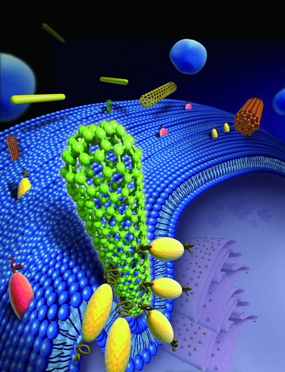
Carbon nanotubes and other long nanomaterials can spell trouble for cells. The reason: Cells mistake them for spheres and try to engulf them. Once they start, cells cannot reverse course, and complete ingestion never occurs. Researchers at Brown University detail for the first time how cells interact with carbon nanotubes, gold nanowires and asbestos fibers. Results are published in Nature Nanotechnology.

PROVIDENCE, R.I.
[Brown University] — It’s been long known that asbestos spells trouble for human cells. Scientists have seen cells stabbed with spiky, long asbestos fibers, and the image is gory: Part of the fiber is protruding from the cell, like a quivering arrow that’s found its mark.


Something perpendicular this way comesCells ingest things by engulfing them. When a long
perpendicular fiber comes near, the cell senses
only its tip, mistakes it for a sphere, and begins
engulfing something too long to handle.
Credit: Gao Lab/Brown University
But scientists had been unable to understand why cells would be interested in asbestos fibers and other materials at the nanoscale that are too long to be fully ingested. Now a group of researchers at Brown University explains what happens. Through molecular simulations and experiments, the team reports in Nature Nanotechnology that certain nanomaterials, such as carbon nanotubes, enter cells tip-first and almost always at a 90-degree angle. The orientation ends up fooling the cell; by taking in the rounded tip first, the cell mistakes the particle for a sphere, rather than a long cylinder. By the time the cell realizes the material is too long to be fully ingested, it’s too late.

“It’s as if we would eat a lollipop that’s longer than us,” said Huajian Gao, professor of engineering at Brown and the paper’s corresponding author. “It would get stuck.”
The research is important because nanomaterials like carbon nanotubes have promise in medicine, such as acting as vehicles to transport drugs to specific cells or to specific locations in the human body. If scientists can fully understand how nanomaterials interact with cells, then they can conceivably design products that help cells rather than harm them.
“If we can fully understand (nanomaterial-cell dynamics), we can make other tubes that can control how cells interact with nanomaterials and not be toxic,” Gao said. “We ultimately want to stop the attraction between the nanotip and the cell.”

Misrecognition
Receptors on the cell’s surface crowd around the nanotube, effectively standing it upright. The cell mistakes the tube for a sphere and begins to engulf it.
 Credit: Gao Lab/Brown University

Like asbestos fibers, commercially available carbon nanotubes and gold nanowires have rounded tips that often range from 10 to 100 nanometers in diameter. Size is important here; the diameter fits well within the cell’s parameters for what it can handle. Brushing up against the nanotube, special proteins called receptors on the cell spring into action, clustering and bending the membrane wall to wrap the cell around the nanotube tip in a sequence that the authors call “tip recognition.” As this occurs, the nanotube is tipped to a 90-degree angle, which reduces the amount of energy needed for the cell to engulf the particle.
Once the engulfing — endocytosis — begins, there is no turning back. Within minutes, the cell senses it can’t fully engulf the nanostructure and essentially dials 911. “At this stage, it’s too late,” Gao said. “It’s in trouble and calls for help, triggering an immune response that can cause repeated inflammation.”
The team hypothesized the interaction using coarse-grained molecular dynamic simulations and capped multiwalled carbon nanotubes. In experiments involving nanotubes and gold nanowires and mouse liver cells and human mesothelial cells, the nanomaterials entered the cells tip-first and at a 90-degree angle about 90 percent of the time, the researchers report.
“We thought the tube was going to lie on the cell membrane to obtain more binding sites. However, our simulations revealed the tube steadily rotating to a high-entry degree, with its tip being fully wrapped,” said Xinghua Shi, first author on the paper who earned his doctorate at Brown and is at the Chinese Academy of Sciences in Beijing. “It is counter-intuitive and is mainly due to the bending energy release as the membrane is wrapping the tube.”
The team would like to study whether nanotubes without rounded tips — or less rigid nanomaterials such as nanoribbons — pose the same dilemma for cells.
“Interestingly, if the rounded tip of a carbon nanotube is cut off (meaning the tube is open and hollow), the tube lies on the cell membrane, instead of entering the cell at a high-degree-angle," Shi said.
Agnes Kane, professor of pathology and laboratory medicine at Brown, is a corresponding author on the paper. Other authors include Annette von dem Bussche from the Department of Pathology and Laboratory Medicine at Brown and Robert Hurt from the Institute for Molecular and Nanoscale Innovation at Brown.
The National Science Foundation, the U.S. Department of Commerce National Institute of Standards and Technology, the National Institute of Environmental Health Sciences Superfund Research Program, and the American Recovery and Reinvestment Act funded the research.
By Richard Lewis
Read More
Posted in gao, nano, nanotubes | No comments

Wednesday, 23 March 2011

Nanomodified surfaces seal leg implants against infection

Posted on 11:20 by Unknown
Researchers at Brown University have created nanoscale surfaces for implanted materials that mimic the contours of natural skin. The surfaces attract skin cells that, over time, are shown to build a natural seal against bacterial invasion. The group also created a molecular chain that allows an implant surface to be covered with skin cell-growing proteins, further accelerating skin growth. Results are published in theJournal of Biomedical Materials Research A.
PROVIDENCE, R.I. [Brown University] — In recent years, researchers have worked to develop more flexible, functional prosthetics for soldiers returning home from battlefields in Afghanistan or Iraq with missing arms or legs. But even new prosthetics have trouble keeping bacteria from entering the body through the space where the device has been implanted.
“You need to close (the area) where the bacteria would enter the body, and that’s where the skin is,” said Thomas Webster, associate professor of engineering and orthopaedics at Brown University.
Webster and a team of researchers at Brown may have come across the right formula to deter bacterial migrants. The group reports two ways in which it modified the surface of titanium leg implants to promote skin cell growth, thereby creating a natural skin layer and sealing the gap where the device has been implanted into the body. The researchers also created a molecular chain to sprinkle skin-growing proteins on the implant to hasten skin growth.
The findings are published in the Journal of Biomedical Materials Research A.
Thomas WebsterAssociate Professor of Engineering and OrthopaedicsThe researchers, including Melanie Zile, a Boston University student who worked in Webster’s lab as part of Brown’s Undergraduate Teaching and Research Awards program, and Sabrina Puckett, who earned her engineering doctorate last May, created two different surfaces at the nanoscale, dimensions less than a billionth of a meter.
In the first approach, the scientists fired an electron beam of titanium coating at the abutment (the piece of the implant that is inserted into the bone), creating a landscape of 20-nanometer mounds. Those mounds imitate the contours of natural skin and trick skin cells into colonizing the surface and growing additional keratinocytes, or skin cells.
Webster knew such a surface, roughened at the nanoscale, worked for regrowing bone cells and cartilage cells, but he was unsure whether it would be successful at growing skin cells. This may be the first time that a nanosurface created this way on titanium has been shown to attract skin cells.
The second approach, called anodization, involved dipping the abutment into hydrofluoric acid and giving it a jolt of electric current. This causes the titanium atoms on the abutment’s surface to scurry about and regather as hollow, tubular structures rising perpendicularly from the abutment’s surface. As with the nanomounds, skin cells quickly colonize the nanotubular surface.
In laboratory (in vitro) tests, the researchers report nearly a doubling of skin cell density on the implant surface; within five days, the keratinocyte density reached the point at which an impermeable skin layer bridging the abutment and the body had been created.
“You definitely have a complete layer of skin,” Webster said. “There’s no more gap for the bacteria to go through.”
To further promote skin cell growth around the implant, Webster’s team looked to FGF-2, a protein secreted by the skin to help other skin cells grow. Simply slathering the abutment with the proteins doesn’t work, as FGF-2 loses its effect when absorbed by the titanium. So the researchers came up with a synthetic molecular chain to bind FGF-2 to the titanium surface, while maintaining the protein’s skin-cell growing ability. Not surprisingly, in vitro tests showed the greatest density of skin cells on abutment surfaces using the nanomodified surfaces and laced with FGF-2. Moreover, the nanomodified surfaces create more surface area for FGF-2 proteins than would be available on traditional implants.
The next step is to perform in vivo studies; if they are successful, human trials could begin, although Webster said that could be years away.
The U.S. Department of Veterans Affairs and the U.S. National Science Foundation funded the research.
Read More
Posted in nano, webster | No comments

Friday, 28 January 2011

Nanotechnology research offers possible solution to common implant complication

Posted on 10:06 by Unknown
Thomas Webster, associate professor of engineering, recently completed a study that demonstrates how medications can be deployed on demand from the surface of bone implants to reduce the prevalence of infection. The medications were administered using polypyrroles, electrically conductive polymer films that coat the implants to hold the drugs and release them when voltage is applied.
Full report online: www.nanowerk.com/spotlight/spotid=19835.php

Nanotechnology research lays the foundation for smart implants
(Nanowerk Spotlight) Imagine intelligent medical implants that can continuously monitor their condition inside the body and autonomously respond to changes such as infection by releasing anti-inflammatory agents. Thanks to nanotechnology, medical research is moving quickly towards this goal.
The market for medical implant devices is huge and growing fast – in the U.S. alone it is estimated to be $23 billion per year and it is expected to grow by about 10% annually for the next few years. Implantable cardioverter defibrillators, cardiac resynchronization therapy devices, pacemakers, tissue and spinal orthopedic implants, hip replacements, phakic intraocular lenses and cosmetic implants will be among the top sellers. Each year in the U.S., almost 500,000 patients receive hip and knee replacements, about the same number need bone reconstruction due to injuries or congenital defects and 16 million Americans loose teeth and may require dental implants.
Implant wear and infections remain the major problem facing the long-term success and survival of these artificial joints. Studies have shown that large amounts of minute wear particles are produced by orthopaedic implants (both metal and plastic), setting into motion a cascade of events that ultimately may result in the disappearance of bone around the implant (osteolysis). This can lead to implant loosening and failure of the artificial joint. Diagnosis if and what is wrong with an implant relies on X-rays or other imaging techniques. The techniques are insensitive, not in real time, and require the patient to go into a hospital. Surgery to replace these failures is more difficult to perform, is more costly, and has a poorer outcome than the original joint replacement surgery.
A new study shows that the use of polypyrrole films as electrically controlled drug release devices on implant surfaces can potentially improve bone implants. By electrodepositing antibiotics or anti-inflammatory drugs in a polymer coating on medical devices, researchers at Brown University demonstrate that such drugs can be released from polypyrrole on demand – by applying a voltage – and control cellular behavior important for orthopedic applications, i.e. inhibit inflammation and kill bacteria.
"Polypyrrole is an intrinsically conductive polymer which can be electrochemically synthesized as a thin film on conductive materials," Thomas J. Webster tells Nanowerk. "Polypyrrole has been studied for various applications such as corrosion protection, electrochemical biosensors, electrode coatings, bioelectronics, solid-state devices and patterned circuits, and, although it has emerged as a promising material with substantial potential for biomedical applications and controlled drug delivery, few studies have investigated its possible role in decreasing infection and inflammation for orthopedic applications."
layered titanate nanosheets layered titanate nanosheets
Left: Polypyrrole electrodeposited on conventional titanium. Right: Polypyrrole electrodeposited on multi-walled carbon nanotubes (Images: Webster Lab, Brown University)
Reporting their findings in the January 17, 2011 online issue of Nanotechnology ("Electrically controlled drug release from nanostructured polypyrrole coated on titanium"), Webster and his team demonstrate a 'proof of concept' to develop and evaluate on-demand delivery of penicillin/streptomycin (antibiotics used to treat grampositive and gram-negative bacteria) and dexamethasone (a glucocorticoid used clinically as an anti-inflammatory and immunosuppressive agent) in situ from polypyrrole. It shows that medications can be deployed on demand from the surface of bone implants to reduce the prevalence of both septic and aseptic complications.
This work is an extension of previous studies from Webster's nanomedicine lab demonstrating that these nanostructured materials sense and promote new bone growth ("Greater osteoblast functions on multiwalled carbon nanotubes grown from anodized nanotubular titanium for orthopedic applications"and "Multiwalled carbon nanotubes enhance electrochemical properties of titanium to determine in situ bone formation").
To create their polymer coating, the researchers first grew multi-walled carbon nanotubes (approx. 55nm in diameter) out of anodized nanotubular titanium using cobalt-catalyzed chemical vapor deposition. Polypyrrole monomers were either oxidized with the antibiotics or the dexamethasone before electrochemical polymerization of the polypyrrole around these nanotubes was carried out.
"Anionic drugs, bound electrostatically inside the polypyrrole thin film, were released in our present study by the application of a negative voltage," explains Webster. "For the first five cycles, we observed the anionic molecules to move in and out of the polypyrrole thin film due to continuous oxidation and reduction. The reduction peaks of penicillin/streptomycin release disappeared after 15 cyclic voltammetry cycles. For dexamethasone, the reduction peak was still observed after 25 cycles, but disappeared after 40 cycles."
The increase in the amount of drugs released after the electrical excitation was significant until 5 cycles. The cumulative release of penicillin/streptomycin and dexamethasone approached 80% of the drug and no further drug release was observed with further cycles.
Webster notes that, although they found that polypyrrole can be overoxidized and lose its electroactivity at higher potentials or when voltages are applied for longer periods, carbon nanotubes can maintain and prolong the electroactivity of polypyrrole due to their excellent conductivity properties.
He points out that polypyrrole can be doped not only with antibiotics and drugs like dexamethasone – allowing the preloading of drugs to obtain clinically controllable and predictable doses – but also various other biomolecules such as for instance growth factors, peptides, enzymes, antibodies, proteins, etc. to alter its biological, physical, chemical and electrical properties to design a controlled released system for numerous biomedical applications.
In addition, polypyrrole can be coated on electrodes or integrated with implantable chips to introduce an electrical signal into the biological environment.
"These preliminary results lay the foundation for developing intelligent orthopedic drug delivery technologies that can utilize a closed-loop sensing process with drug administration based on that sensing information," says Webster. "Both carbon nanotube-based sensors and controllable drug delivery systems could be an excellent way to improve the lifetime of orthopedic implants, allowing implants to kill bacteria, reduce the susceptibility of implants to prolonged inflammatory responses and ultimately increase bone formation."
By Michael Berger. Copyright 2011 Nanowerk
Read More
Posted in implant, nano, webster | No comments
Older Posts Home
Subscribe to: Posts (Atom)

Popular Posts

  • Clyde Briant elected to the National Academy of Engineering
    Congratulations to Clyde Briant on his election to the National Academy of Engineering. Election to the National Academy of Engineering is ...
  • Nanoparticles eliminate bacterial infection on prosthetic
    Infected implants now have a foe. Brown University researchers have created a nanoparticle that can penetrate a bacterial-produced film on p...
  • Hydrodynamics of Microorganisms
    Professor Powers' article, Hydrodynamics of Swimming Mircoorganisms, was accepted this week in Reports on Progress in Physics. Preview t...
  • AIChE Prize Winners
    This year, in celebration of AIChE's centennial, the AIChE Annual Meeting featured special sessions that highlight chemical engineering...
  • RI Woman of the Year
    The Rhode Island Commission on Women (RICW) is pleased to announce the 2009 Women of the Year, Lynn Ewart-Paine, ’85 ’86 Sc.M. ’90 Ph.D., S...
  • Nanosatellite Launch Vehicle Conference
    In the over 25 years since small, low cost space began its transition from curiosity to the integral element of world-wide civil and militar...
  • NanoVis, Inc earns 2nd in Venture Idol
    NanoVis, Inc., a Brown start-up company, Gets 2nd Place in Venture Idol Competition NanoVis, Inc., a Brown start-up company was awarded 2nd ...
  • Superfund renewed to create a safer and healthier RI
    Brown's Superfund Research Program (SRP) brings together researchers from engineering, biology and medicine, geological sciences, and so...
  • IGEM Jamboree
    The 2008 Brown iGEM Teams worked on two projects--one a toxin detection & electrical reporting system using E. Coli bacteria and the se...
  • Wei Yang to lead China’s National Natural Sciences Foundation
    Wei Yang Ph.D.’85 Sc.D.’12 hon., an internationally celebrated engineer and materials science researcher, educator, and administrator, has ...

Categories

  • "Sirinrath Sirivisoot"
  • aaas
  • aaron
  • abet
  • accreditation
  • aceros
  • ACS
  • admission
  • admissions
  • advanced baby imaging lab
  • AIChE
  • AIChE ugrad award
  • AIMBE
  • almeida
  • alumni
  • Andrew Peterson
  • apoE4
  • archaeology
  • archambault
  • argyria
  • ARPA-E
  • article
  • artificial ovary
  • asme
  • associate dean
  • audax
  • audio
  • awar
  • award
  • axena
  • Aziz
  • Baek
  • bahar computer conference
  • banyan
  • bashevkin
  • bats
  • battery
  • beam
  • BEAR Day
  • bio
  • biodiesel
  • biofilm
  • biofuel
  • blume
  • bme
  • bmes
  • book
  • borton
  • bower
  • brain initiative
  • Brain Science
  • brain sensor
  • brain-computer interface
  • braingate
  • braingate2
  • breuer
  • breuer bats reuters
  • briant
  • brown institute of brain science
  • bull
  • bull risd car
  • business plan
  • calakli
  • calo
  • CAMR
  • car
  • carbon
  • career fair
  • Caswell
  • catena
  • cave
  • CfNN
  • chemical innovation program
  • China
  • civil engineering
  • coda
  • coe-sullivan
  • collaboration
  • commencement
  • competition
  • Computational Materials Science
  • concussion
  • cooper
  • cord-clamping
  • crisco
  • crisco risd
  • CRL
  • Curet
  • curtin
  • cyberkinetics
  • dang
  • dean
  • decker
  • deisley
  • deoni
  • desai
  • desktop delta-v
  • dingman
  • DOE
  • donoghue
  • donovan
  • Durmus
  • dworak
  • eastman conference
  • ejiofor
  • election
  • elevator pitch
  • emanuel
  • emotive
  • Empower
  • en4
  • energy-momentum spectroscopy
  • ENGN 1930G
  • entrepreneurship
  • Entreprenuership
  • EPSCOR
  • escuti
  • event
  • ewb
  • faculty
  • feature
  • fellowship
  • Felzenszwalb
  • fleeter
  • fluid dynamics
  • franck
  • freund
  • fsae
  • fulbright
  • gao
  • General Motors
  • GhostBot
  • gidmark
  • gingerbread
  • GM
  • GM/Brown
  • grad
  • graduate
  • grant
  • grantab
  • graphene
  • greis
  • guduru
  • guo
  • haberstroh
  • halpin
  • halpin prize
  • Hargus
  • hazeltine
  • heart
  • hibbitt
  • HnC
  • hochberg
  • huebscher
  • hurt
  • hurt cfl nyt
  • hydrokinetic
  • IE
  • IIT-Bombay
  • IMNI
  • implant
  • Indo-US
  • innovation
  • institute of medicine
  • international
  • internship
  • jackson
  • jadhav
  • jakubek
  • Jay
  • JCD Wind
  • jepsen
  • joukowsky
  • JPL
  • kane
  • kesari
  • keynote
  • Kim
  • klout
  • Kristie Chin
  • kulaots
  • Külaots
  • kumar
  • Kummer
  • lacrosse
  • larson
  • laser
  • laulicht
  • lavernia
  • Lazos
  • Lee
  • LEGO
  • LIB
  • light emission
  • liquid bone
  • lithium ion battery
  • Liu Finalist GEMS Award
  • lubricin
  • lysaght
  • magnet
  • malik
  • Mandre
  • maris
  • materials
  • Materials Research
  • mathiowitz
  • mba
  • mccalla
  • Mechanics
  • Megan Buczynski
  • mentor
  • mercury
  • metamaterials
  • metaphotonics
  • MGI
  • mittlemann
  • morgan
  • muri
  • NAE
  • nano
  • nanoparticles
  • nanopatch
  • nanoscience
  • nanoskin
  • nanotechnology
  • nanotubes
  • nanovis
  • NASA
  • Needleman
  • neuroengineering
  • Neurorestoration
  • Neuroscience
  • neurotechnology
  • NewMech
  • NewMech2012
  • nih
  • nsf
  • NSFC
  • nurmikko
  • nurse
  • open house
  • optical
  • osteoarthritis
  • overhead.fm
  • pacifici
  • padture
  • palmore
  • palmore hoffmankim nih
  • paper
  • patent
  • paxson
  • pecase
  • Peterson
  • petteruti
  • Phi Beta Kappa
  • photos graduation
  • powers
  • powers editor journal
  • president
  • prime
  • PRIME Omega-3
  • Privicare
  • PriWater
  • profiles
  • project
  • publication
  • publication leadership
  • qd vision
  • Raimondo
  • rainwater
  • ramesh
  • Ramos
  • rankings
  • reda
  • reed
  • Reggiannini
  • research
  • richardson
  • risd
  • Riviere
  • robot
  • robots
  • rome
  • rosakis
  • Rosenstein
  • Runa
  • salomon award
  • sarin
  • schutter
  • scripta materialia
  • selenium
  • SES
  • sharp
  • sheldon
  • shenoy
  • sigma xi
  • silver
  • Silverman
  • simeral
  • simulia
  • SMART
  • solar
  • Solar4Cents
  • space
  • Speramus
  • Spira
  • stac
  • startup
  • stem outreach
  • Stout
  • summer
  • superfund
  • suuberg
  • swe
  • sygiel
  • takamoto biogas
  • tau beta pi
  • taubin
  • taylor
  • team
  • timoshenko
  • tissue
  • tissue engineering
  • tran
  • tripathi
  • tsang
  • twitter
  • ugrad
  • VA
  • van de Walle
  • venture for america
  • video
  • Vlahovska
  • wadia
  • wang
  • warshay
  • watson
  • website
  • webster
  • webster nano
  • webster nurmikko bio conference
  • Wells
  • weng
  • wireless
  • workshop
  • yang
  • yin
  • zhang
  • zhang webster star award phd
  • zia
  • zia nsf award

Blog Archive

  • ▼  2013 (18)
    • ▼  April (1)
      • Nurmikko and Donoghue join U.S. BRAIN initiative
    • ►  March (5)
    • ►  February (4)
    • ►  January (8)
  • ►  2012 (76)
    • ►  December (5)
    • ►  November (8)
    • ►  October (9)
    • ►  September (5)
    • ►  August (6)
    • ►  July (6)
    • ►  June (5)
    • ►  May (4)
    • ►  April (8)
    • ►  March (11)
    • ►  February (6)
    • ►  January (3)
  • ►  2011 (95)
    • ►  December (7)
    • ►  November (9)
    • ►  October (8)
    • ►  September (11)
    • ►  August (8)
    • ►  July (4)
    • ►  June (3)
    • ►  May (10)
    • ►  April (7)
    • ►  March (10)
    • ►  February (10)
    • ►  January (8)
  • ►  2010 (55)
    • ►  December (13)
    • ►  November (8)
    • ►  October (3)
    • ►  September (2)
    • ►  August (5)
    • ►  July (1)
    • ►  June (2)
    • ►  May (8)
    • ►  April (5)
    • ►  March (2)
    • ►  February (4)
    • ►  January (2)
  • ►  2009 (46)
    • ►  December (7)
    • ►  November (2)
    • ►  October (4)
    • ►  September (4)
    • ►  August (2)
    • ►  July (5)
    • ►  June (4)
    • ►  May (6)
    • ►  April (5)
    • ►  March (1)
    • ►  February (4)
    • ►  January (2)
  • ►  2008 (15)
    • ►  December (1)
    • ►  November (4)
    • ►  October (1)
    • ►  September (1)
    • ►  August (2)
    • ►  July (3)
    • ►  June (2)
    • ►  April (1)
  • ►  2007 (1)
    • ►  February (1)
Powered by Blogger.

About Me

Unknown
View my complete profile